VV ECMO Part 3 of 3: Two Strategies to Transition to Ventilator-Only Support
As a novice ECMO specialist, I am discovering the complexities and nuances of managing and weaning patients from VV ECMO. This article is part three of a three-part series, reflecting my ongoing learning journey. In this article, we will discuss the weaning process from VV ECMO and a couple of special considerations during weaning. There are many additional elements to consider for a successful wean, such as fluid management, sedation, physical therapy, and anticoagulation, which will be discussed in future articles.
Why Use VV ECMO?
Venovenous ECMO (VV ECMO) is primarily utilized for patients with severe respiratory failure unresponsive to conventional therapies. This form of ECMO becomes necessary in several critical scenarios:
-
Refractory Respiratory Failure: VV ECMO is employed when severe respiratory failure does not improve with standard treatments such as mechanical ventilation, prone positioning, and pharmacological interventions. This includes cases where hypoxemia and/or hypercapnia persist despite maximal medical support.
-
High Ventilator Pressures: VV ECMO can provide the necessary gas exchange support when maintaining adequate oxygenation and ventilation requires ventilator settings that exceed safe parameters, such as very high positive end-expiratory pressure (PEEP) or peak inspiratory pressures. Please look at a previous article showing that excessively high ventilator pressures can cause ventilator-induced lung injury (VILI), barotrauma, or volutrauma. VV ECMO allows for lung-protective ventilation strategies, reducing the risk of further lung damage by enabling lower ventilator pressures and tidal volumes.
Lung Transplantation:
-
Pre-Transplant: For patients awaiting lung transplantation, it is crucial to minimize ventilator-induced injury and maintain optimal oxygenation and ventilation. VV ECMO provides a safe method to achieve these goals, ensuring adequate gas exchange while using lower ventilator pressures. This approach helps to preserve the patient’s lung function and overall condition, making them a better candidate for successful transplantation.
-
Post-Transplant: After lung transplantation, VV ECMO can be essential for patients with fragile surgical sites. The newly transplanted lungs may require support to optimize healing and function without the stress of high ventilator pressures. VV ECMO helps to stabilize the patient and provides adequate oxygenation and carbon dioxide removal, which are critical for the recovery of the delicate transplanted tissues.
Specific Conditions:
-
Acute Respiratory Distress Syndrome (ARDS): Severe ARDS, particularly when caused by conditions such as sepsis, trauma, or viral infections like influenza and COVID-19, can result in refractory hypoxemia.
-
Severe Pneumonia: In severe pneumonia, conventional mechanical ventilation fails to maintain adequate gas exchange.
-
Severe Asthma: Life-threatening asthma attacks that do not respond to standard treatments can lead to respiratory failure, requiring ECMO support.
-
Massive Pulmonary Embolism: When a large pulmonary embolism severely impairs gas exchange, and conventional therapies are insufficient.
In summary, VV ECMO is a critical intervention for patients who require an alternative method to achieve adequate oxygenation and ventilation, especially when conventional ventilation strategies pose a risk of further lung injury, are inadequate for maintaining life-sustaining gas exchange, or when managing pre- and post-lung transplantation cases with fragile surgical sites. For a deeper dive, you can read one of my previous articles, VV ECMO Part 1 of 3: Indications and Rationale for Use.
Weaning from VV ECMO
Weaning from venovenous (VV) ECMO begins by recognizing lung recovery and decreasing the oxygenation and CO2 removal provided by the ECMO circuit as the patient’s lung function improves. The process involves setting the ventilator to acceptable levels (e.g., Pplat < 30 cm H2O, PEEP 8-12 cm H2O, FiO2 < 0.5-0.6) and then discontinuing the sweep gas flow across the oxygenator while continuing blood flow. The sweep gas can be discontinued by simply turning off the flow. Allowing room air to flow across the membrane lung can provide ongoing gas transfer.
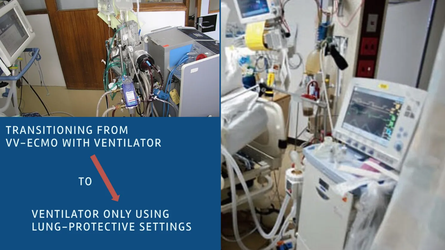
The patient’s underlying condition, which led to ECMO, must have improved. The weaning process is considered diagnostic rather than therapeutic, as it helps determine whether the patient can breathe independently. Image from Wikimedia Commons, public domain
Note
Weaning Strategies for VV ECMO
Strategy 1: Using the Blender to Reduce FDO2
-
Assessment of Lung Function: Regular evaluations of ABGs, chest X-rays, and clinical status. Ensure resolution or improvement of the underlying cause of respiratory failure.
-
Initial Reduction of Sweep Gas Flow: Gradually decrease sweep gas flow from higher levels (e.g., 8-10 LPM) to lower levels. Monitor ABGs to ensure CO2 removal and prevent acidosis.
-
Reduction of FDO2 Using Blender: Once sweep gas flow is stable at a low level (e.g., 2-3 LPM), gradually reduce FDO2 using the blender. Decrease FDO2 in small increments (e.g., 0.1 or 0.05) while monitoring ABGs and SpO2 to ensure adequate oxygenation.
-
Ongoing Monitoring: Continuously monitor respiratory status, lung compliance, and hemodynamics. Please be sure to watch for signs of respiratory distress or instability.
-
Final Trial Off ECMO: Conduct a trial off ECMO by clamping the circuit and monitoring the patient’s gas exchange. If the patient maintains adequate oxygenation and ventilation, proceed to decannulation.
-
Decannulation: Remove ECMO cannulas and provide post-decannulation care, including respiratory support.
Strategy 2: Not Using the Blender (Leaving FDO2 at 1.00)
-
Assessment of Lung Function: Regular evaluations of ABGs, chest X-rays, and clinical status. Ensure resolution or improvement of the underlying cause of respiratory failure.
-
Gradual Reduction of Sweep Gas Flow: Gradually decrease sweep gas flow from higher levels (e.g., 8-10 LPM) to below 1 LPM while keeping FDO2 at 1.00. Monitor ABGs to ensure CO2 removal and prevent acidosis.
-
Monitoring CO2 Clearance: Gradually decrease sweep gas flow from higher levels (e.g., 8-10 LPM) to below 1 LPM while keeping FDO2 at 1.00. Monitor ABGs to ensure CO2 removal and prevent acidosis.
-
Automatic Adjustment of Oxygenation: As the sweep gas flow decreases below 1 LPM, the SaO2, and pO2 may naturally start to reduce due to the efficiency of the oxygenator. The reduced contact time between blood and the gas in the membrane lung results in lower oxygen transfer, effectively mimicking a reduction in FDO2. The blood flow to sweep gas flow ratio increases, contributing to this effect.
-
Further Reduction of Sweep Gas Flow: Decrease sweep gas flow to as low as possible (e.g., 0.5 LPM or less). Ensure stable CO2 clearance and oxygenation through frequent ABG analysis.
-
Final Trial Off ECMO: Conduct a trial off ECMO by clamping the sweep gas and monitoring the patient’s gas exchange. If the patient maintains adequate oxygenation and ventilation, proceed to decannulation.
-
Decannulation: Remove ECMO cannulas and provide post-decannulation care, including respiratory support.
Ventilator Management During Weaning
During either weaning strategy, it is crucial to keep ventilator settings within safe parameters:
-
FiO2: Maintain FiO2 between 30-60% to avoid oxygen toxicity.
-
PEEP: Maintain PEEP at 5-10 cmH2O to support alveolar recruitment and prevent atelectasis.
-
Tidal Volume (Vt): For volume control mode, increase Vt in increments of 1-2 mL/kg of predicted body weight (PBW) to a maximum of 6-8 mL/kg of PBW with acceptable plateau pressures (Pplat ≤ 28 cmH2O).
-
Driving Pressure: For pressure control or pressure support modes, increase the driving pressure to no more than 15-20 cmH2O with delivered Vt in the 6-8 mL/kg of PBW range.
-
Respiratory Rate (RR): Keep RR ≤ 30 breaths/min.
The goal is to ensure lung protective ventilation while decreasing ECMO support, allowing the patient’s lungs to recover and resume their function independently. If higher ventilator settings are required, this indicates that the patient may not be ready for weaning off ECMO.
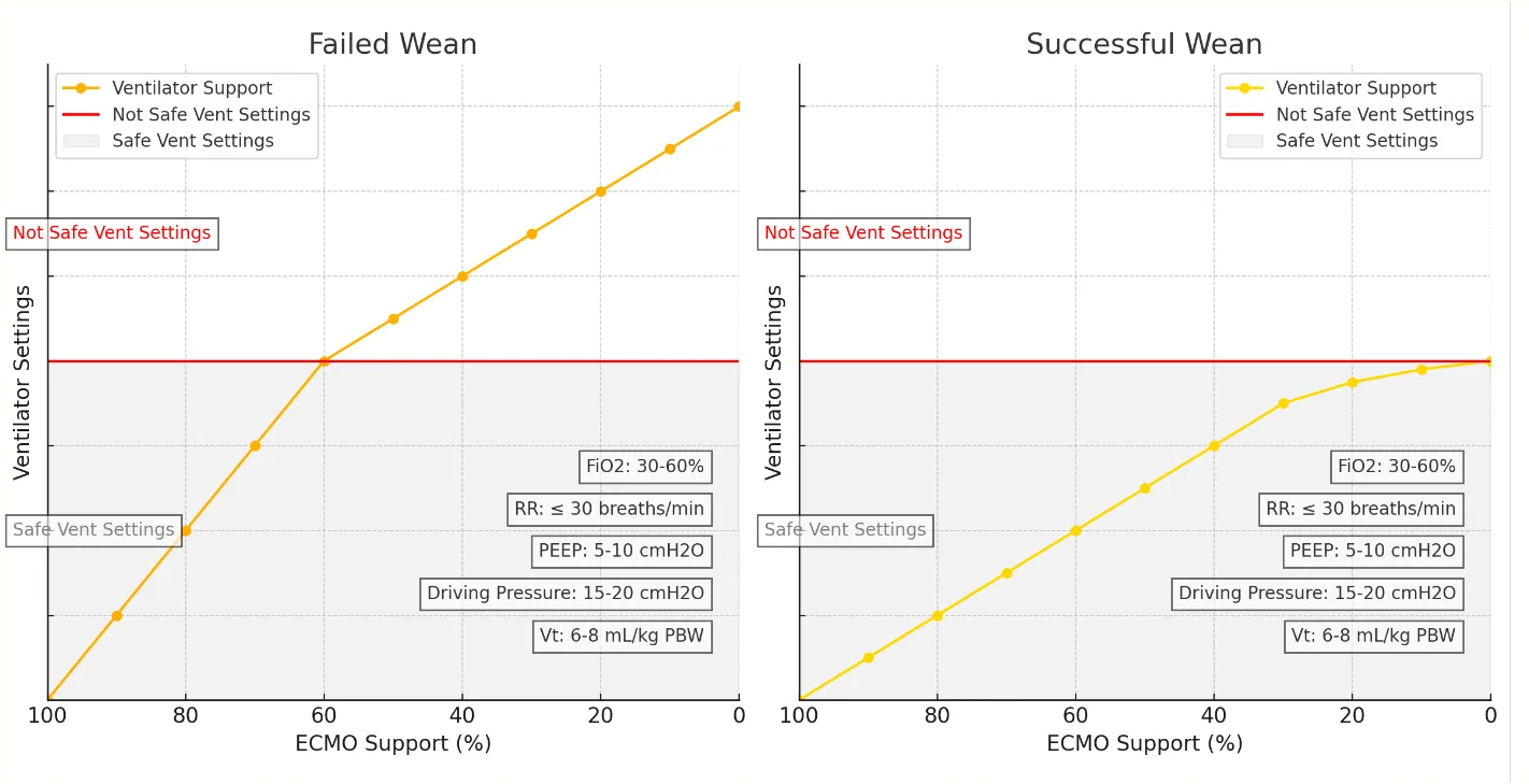
Above the red line indicates failed weaning, where ventilator settings no longer protect the lungs. At this point, weaning from VV ECMO would cease, and higher ECMO settings would be used to revert the ventilator to lung-protective settings. Weaning would be attempted again later to allow the lungs time to heal. Charts generated using OpenAI
Special Considerations after Sweep Gas is Disconnected
- Extended Trial-Off: Some patients are marginal during the usual 2-hour trial-off procedure, but the team may feel that ventilation or PA pressure may become acceptable with time for pulmonary toilet, ventilation adjustment, sedation, and gentle lung recruitment. The trial-off can be extended until acceptable blood gases and PA pressures are obtained in lung-protective ventilator settings. It is unusual to extend a VV trial-off beyond 24 hours. If performing a prolonged VV trial-off, reduce blood gas frequency stepwise to 6-hourly as the trial progresses.
Additional Considerations for Successful Weaning
Weaning a patient from VV ECMO involves more than just adjusting the ECMO parameters. A successful wean requires a comprehensive approach that includes:
-
Fluid Management: Carefully manage fluid balance to prevent fluid overload while ensuring adequate perfusion and organ function.
-
Sedation: Adjust sedation to allow for patient comfort and cooperation, minimizing sedation as the patient’s condition allows for assessing neurological status and facilitating weaning.
-
Physical Therapy: Engage in early mobilization and physical therapy to enhance respiratory muscle strength and overall physical recovery.
-
Anticoagulation Management: Monitor and adjust anticoagulation therapy to prevent thromboembolic events while avoiding bleeding complications.
These considerations are very important for a successful wean, but I will not discuss these additional points in this article. I will delve into these other critical areas in a future article.
Conclusion
Venovenous ECMO (VV ECMO) is a crucial intervention for patients with severe respiratory failure who are unresponsive to conventional therapies. It provides a life-saving method to maintain adequate oxygenation and ventilation, particularly in scenarios where high ventilator pressures are required or in the context of lung transplantation with fragile surgical sites. Employing VV ECMO helps minimize ventilator-induced lung injury and allows for lung-protective ventilation strategies, essential for the recovery and preservation of lung function.
References
-
Extracorporeal Life Support: The ELSO Red Book, 6th Edition. Published by the Extracorporeal Life Support Organization (ELSO), this comprehensive text is a key resource for understanding the principles and practices of ECMO.
-
ECMO Specialist Training Manual, 4th Edition. This manual provides in-depth training protocols and guidelines for ECMO specialists, covering cannulation techniques, patient management, and emergency procedures.
-
Various Other Source Materials. Additional insights and guidelines on ECMO practice, including hospital patient care practices.
Note: This article reflects my learning journey in ECMO and is intended for educational purposes only. It should not be used as a substitute for professional medical advice or guidance. Always consult with qualified healthcare professionals for clinical decisions and patient care.
Share and Subscribe. Join the ECMO 143 Learning Journey
Other Links:
Follow me on LinkedIn: Jonathan B. Jung, RRT-NPS
Follow me on X (Twitter) “ECMO 143-Stay Uptodate” List on X
Acknowledgments:
I developed three custom GPTs, “AI ECMO Expert,” “ECMO Specialist Handover Practice,” and “Micro Definitions (MD-GPT),” for specialized research. These tools draw primarily from the ELSO Redbook (6th Edition), the ELSO Specialist Training Manual (4th Edition), various research papers, and articles. Additional research was supported by GPT-4o/o1, Claude 3.5 Sonnet/Opus, and Perplexity. Editing was performed with Grammarly. A.I. images and charts were created using Leonardo AI, DALL-E3 AI Image Generator, Microsoft Designer, and Adobe Express. Content for all articles sourced from Extracorporeal Life Support: The ELSO Red Book, 6th Edition, and ECMO Specialist Training Manual, 4th Edition.